“Back trouble” factoids….

Inheritability of back pain may be as high as 68% with inheritable factors including disc degeneration (statistically accounting for up to 75%) and tendency toward psychological distress.  Four studies draw the conclusion muscle strength and general fitness appear to be of little significance and there is no persuasive evidence flexibility, gender or leg length inequality play a meaningful role. Taller men do have a higher prevalence of certain disc problems presumable due to the fact tall people lift weights at the end of longer lever arms. Several studies have concluded there is “moderate evidence” that lumbar flexibility, bodyweight and straight leg raise have no predictive value for future LBP. Additionally recent studies also show moderate evidence that general cardiorespiratory fitness has no predictive value for future LBP.  And has been mentioned in other blogs MRI and radiographs hold NO value in predicting future LBP.

A relatively recent systematic review (Pincus et al Spine 2002) of psychological factors as predictors of “unfavorable outcomes” concluded there is “good evidence” to implicate distress/depressive mood and somatization in development of chronicity.

And there is also “strong evidence” psychological factors play an important role in persisting symptoms. A workers’ belief regarding their work having caused their pain and their expectations of inability to return (‘yellow flags’) are of particular importance.

Revisiting MRI findings: circa 1990 (Bodon et al Mar J Bone Joint Surg)

They found 1/3rd of asymptomatic individuals to have a “substantial abnormality”.
Those under 60; 25% had a HNP, over 60 nearly 60% showed abnormalities; 40% HNP and 25% stenosis. Degeneration and bulging was found in more than a 1/3rd of those under 40 and in everyone 60 and older.
In 1994 similar findings were demonstrated in a study in the New England Journal of Medicine.
MRI T2 weighted images reveal that degeneration may begin in the teens especially in athletic individuals.
Degenerated discs are ubiquitous and assumed a source of pain but how they are “turned on” remains elusive…however an untenable or uncontrolled torque is usually the mechanical act that may initiates the pain (dietary and psychological factors notwithstanding).
I will guess that eventually MR scans (or another technology) will become so detailed as to reveal the microscopic fibers and the “active tear” where the pain probably generates. Why the pain fails to disappear after months in 10-15% is also elusive but recurrent mechanical stress and inefficient systemic healing must play a role.

Check out these 2 studies on ‘nodules’ and back pain.

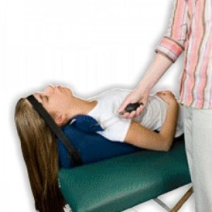
I came upon 2 studies recently which really piqued my interest immediately. The suggestion by many patients that the mobile “lump”/nodule in their lower back is “causing” their trouble is VERY common in my experience. These two commentaries point out that in fact the patient may not just be imagining that the proximity of the lump to their pain isn’t coincidence…the nodule may well be the source.  I have encountered several notable cases of this over the last 28 years, but recently had a post-surgical patient absolutely convinced that “the damn lump in my back is giving me the pain!”…”but no one will believe me”. After probing the area it became apparent that it wasn’t just an “overlaying” lump (common with many lipomas which are not a pain source), this lump appeared to be the actual source of the pain. the pain interestingly enough was a “discal” like pain but with nebulous motion-findings. The lump was injected, pain relief was dramatic and it was removed. And the patient was extremely satisfied with the outcome.

Aqri 2013;25(2):83-6. doi: 10.5505/agri.2013.63626.

[Episacral lipoma: a treatable cause of low back pain].

[Article in Turkish]

Erdem HR1Nacır BÖzeri ZKaragöz A.


Abstract

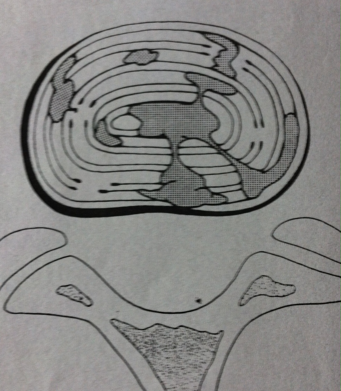
Episacral lipoma is a small, tender subcutaneous nodule primarily occurring over the posterior iliac crest. Episacral lipoma is a significant and treatable cause of acute and chronic low back pain. Episacral lipoma occurs as a result of tears in the thoracodorsal fascia and subsequent herniation of a portion of the underlying dorsal fat pad through the tear. This clinical entity is common, and recognition is simple. The presence of a painful nodule with disappearance of pain after injection with anaesthetic, is diagnostic. Medication and physical therapy may not be effective. Local injection of the nodule with a solution of anaesthetic and steroid is effective in treating the episacral lipoma. Here we describe 2 patients with painful nodules over the posterior iliac crest. One patient complained of severe lower back pain radiating to the left lower extremity and this patient subsequently underwent disc operation. The other patient had been treated for greater trochanteric pain syndrome. In both patients, symptoms appeared to be relieved by local injection of anaesthetic and steroid. Episacral lipoma should be considered during diagnostic workup and in differential diagnosis of acute and chronic low back pain. 2000 Apr;49(4):345-8.

Fibro-fatty nodules and low back pain. The back mouse masquerade.

Curtis P1Gibbons G

Abstract

BACKGROUND:

Few useful interventions exist for patients with persistent low back pain. We suggest that a fibro-fatty nodule (“back mouse”) may be an identifiable and treatable cause of this and other types of pain.

METHODS:

We describe 2 patients with painful nodules in the lower back and lateral iliac crest areas. In both cases, the signs and symptoms were unusual and presented at locations distant from the nodule. One patient complained of severe acute lower abdominal pain, and the other had been treated for chronic recurrent trochanteric bursitis for several years.

RESULTS:

In both patients, symptoms appeared to be relieved by multiple injection of the nodule.

DISCUSSION:

There is agreement that back mice exist. Referred pain from the nodules might explain the distant symptoms and signs in these cases. Multiple puncture may be an effective treatment because it lessens the tension of a fibro-fatty nodule.

CONCLUSIONS:

Randomized trials on this subject are needed. In the meantime, physicians should keep back mice in mind when presented with atypical and unaccountable symptoms in the lower abdomen, inguinal region, or legs.


The Kdt “bolster-less” leg support feature

Before traction can be transmitted to the spine, pelvic anterior rotation (and lordosis) must be reduced. The lumbar lordosis follows from the reduction of the anterior rotation of the pelvis.

On the Kdt Neural-flex we incorporated the leg and hip support (creating semi-fowler positioning) into the “bolster-less” leg feature. This eliminates the need for a separate tilting-piece and the clumsy and cumbersome knee bolsters…which offer zero variability.

The table-section however allows a near infinite degree of elevation and tilt. Some patients and conditions require greater elevation others less. The extent of leg/hip elevation is most typically based on patient comfort and convention i.e. approximately 30° for most. As to objective indicators there aren’t any absolutes. We recommend the table remain fully “down” for most supine treatments, but elevation will change the angle of incidence (as will the leg support section) so “playing” with both can help locate an optimum pull vector. In 1997 the DRS introduced the concept of “targeting” discs…L5 was 10°, L2/3 30° etc. Though not technically possible, IF a less than optimum response is seen with the typical 30° pull, table height and leg-elevation changes can often cater the pull more to the individual and potentially improve the outcome. However knowing the perfect angle prior to the treatment is doubtful.

“Self-decompression” & the seated chair lift

seated chair liftDon’t underestimate the value of seated ‘chair-lifts’ both as an important testing procedure for “compression but also as a counter to the discal stress imposed by daily sitting. Holtzman, Froyer, Saunders and others have examined the decompression effect and benefit of this maneuver and found that it indeed both relieves pain and restores height i.e. fosters disc fluid imbibition in people who sit a lot during the day. “Sitting is the new smoking” takes on a chiropractic edge when you realize the damaging effects on the lumbar discs. Instructing patients to try this “self-decompression” 30-60 seconds (dependent on their strength) several times per day can help pain and possibly disc nutrition & revitalization.

Therapeutic evaluation of lumbar tender point deep massage for chronic non-specific low back pain.

J Tradit Chin Med. 2012 Dec;32(4):534-7.

Zheng Z1Wang JGao QHou JMa LJiang CChen G.


RESULTS:

Patients in the treatment group received tender point deep tissue massage plus lumbar traction and patients in the control group received lumbar traction, alone. We used a tissue hardness meter/algometry and visual analog scale (VAS) to assess the pressure pain threshold, muscle hardness and pain intensity.

CONCLUSION:

Lumbar tender point deep tissue massage combined with lumbar traction produced better improvement in pressure pain threshold, muscle hardness and pain intensity in patients with chronic non-specific low back pain than with lumbar traction alone.

Dr. Kennedy comment: I (and many others) have adopted a lot of ’hands-on’ techniques with decompression since the advent of the “Open” and now Versa-Belt. Ostensibly directed-vertebral contact with an acute HNP or nerve encroachment condition may be counterproductive…however since 90% of presentations are typically less severe hands-on methods during traction can add both clinical benefit and an excellent “selling proposition”. I typically “strip” or deep-pressure the muscles during the rest-phase and let the patient feed-back be my primary guide. Manual oscillations can also be effective.


The association between cervical spine curvature and neck pain

Eur Spine J. May 2007; 16(5): 669–678.

  1. GrobH. Frauenfelder, and A. F. Mannion

Abstract

The finding that degenerative changes of the cervical spine are common in asymptomatic individuals has challenged the notion of cause and effect. Degenerative changes of the cervical spine are commonly accompanied by a reduction or loss of the segmental or global lordosis, and are often considered to be a cause of neck pain. The aim of this study was to examine the correlation between the presence of neck pain and alterations of the normal cervical lordosis in people aged over 45 years. No significant difference between the two groups could be found in relation to the global curvature, the segmental angles, or the incidence of straight-spine or kyphotic deformity (P > 0.05). The presence of such structural abnormalities in the patient with neck pain must be considered coincidental.Degenerative changes of the cervical spine are commonly accompanied by a reduction or loss of the segmental or global lordosis, and are often considered to be a cause of neck pain. Nonetheless, such changes may also remain clinically silent. The aim of this study was to examine the correlation between the presence of neck pain and alterations of the normal cervical lordosis in people aged over 45 years. One hundred and seven volunteers, who were otherwise undergoing treatment for lower extremity problems in our hospital, took part. Sagittal radiographs of the cervical spine were taken and a questionnaire was completed, enquiring about neck pain and disability in the last 12 months. Based on the latter, subjects were divided into a group with neck pain (N = 54) and a group without neck pain (N = 53). The global curvature of the cervical spine (C2–C7) and each segmental angle were measured from the radiographs, using the posterior tangent method, and examined in relation to neck complaints. No significant difference between the two groups could be found in relation to the global curvature, the segmental angles, or the incidence of straight-spine or kyphotic deformity (P > 0.05). Twenty-three per cent of the people with neck pain and 17% of those without neck pain showed a segmental kyphosis deformity of more than 4° in at least one segment—most frequently at C4/5, closely followed by C5/6 and C3/4. The average segmental angle at the kyphotic level was 6.5° in the pain group and 6.3° in the group without pain, with a range of 5–10° in each group. In the group with neck pain, there was no association between any of the clinical characteristics (duration, frequency, intensity of pain; radiating pain; sensory/motor disturbances; disability; healthcare utilisation) and either global cervical curvature or segmental angles. The presence of such structural abnormalities in the patient with neck pain must be considered coincidental, i.e. not necessarily indicative of the cause of pain. This should be given due consideration in the differential diagnosis of patients with neck pain.


Dr. Kennedy comment: another study questioning the ‘curve’ and it’s relation to symptoms. I call attention again to Dr. Eyal  Ledermans’ article: “The fall of the postural-structural-biomechanical model”. However, irrespective of these findings I still say all I’d rather have a ‘good’ curve in my own neck.


Does the sagittal alignment of the cervical spine have an impact on disk degeneration? Minimum 10-year follow-up of asymptomatic volunteers

Eur Spine J. Nov 2009; 18(11): 1644–1651.PMCID:

Eijiro Okada,1et al

Progression of posterior disk protrusion was significantly more frequent in subjects over 40 years of age with non-lordosis type of sagittal alignment. However, no significant correlation between the sagittal alignment of the cervical spine and clinical symptoms was detected. Sagittal alignment of the cervical spine had some impact on the progression of degenerative changes of the cervical spine with aging; however, it had no correlation with the occurrence of future clinical symptoms.

Dr. Kennedy comment:  One of several studies & Meta-analysis of the cervical ‘curve’.  Most are consistent in the determination the curve doesn’t appear to be directly correlated to pain, disability or future prognosis (but this population were asymptomatic, other studies (Gore 1989) suggest only 10% of kyphotic necks are pain-free).  And non-lordosis configurations were related to the most serious protrusions……So(?) that means something!

 The Neck Deck is our inexpensive offering to help address the cervical sagittal alignment. If a patient has persistent neck pain forward head posture, loss of curve, tension myofascial effects and inefficient contraction patterns there should be a strategy to address each of them.

Neck Deck

Revisiting disc degeneration, “migration” & stress shielding

Through 3.5 million years of development we humans find ourselves with lordotic curves intra-uterine. Clearly someone wanted our lower backs and necks “curved” i.e. extended. Numerous published studies suggest extension is the most common directional-preference in HNP conditions and McGill and others show how extension “shuts off” the LE activity thus reducing the crushing-effect of the hyperactive/hyper-tonic posterior muscles endemic in so many with cLBP (Janda’s lower cross syndrome). Further at least 2 recently published studies (Mustafa) show HNP can be effectively treated using extension traction methods. Magnusson, Pope et al (1995)also demonstrated that prone extension postures restore height and have an “unloading” effect on discs equal to or better than flexion postures.

Both a ‘stress-shielding’ (neural arch impaction reducing posterior disc compression) and nuclear “migration” (an intact, hydrophilic nucleus that moves as an isotropic mechanism under pressure) can explain extensions benefit…. but neither is definitive in all cases. It’s not surprising however that extension appears most immediately beneficial in cases of moderate disc degeneration (the arches are already approximating).

So we try to exploit this with our decompression positions…when an extension benefit is apparent prone traction becomes a necessity…when it’s not apparent (or neural stress signs/stenosis or an “anterior disc” manifests) supine is probably the best call.

IDD and its implications to treatment

idd therapy

The discogram shows the effects of IDD (internal disc disruption) and clearly demonstrates why a desiccated and internally degraded nucleus will NOT give “normal/typical” migration-bending effects e.g. flexion & extension findings are ‘inconsistent’.
Since IDD is very common and it’s adverse effects on the hydraulic nature of the disc create a higher potential for annular tears (intolerance to torque) its important to understand its implications to treatment.
Though by no stretch entirely age-dependent it is more likely later in the 4th & 5th decades and suggests why repetitive extensions (facilitated movements) can be counterproductive and prone/prone extended decompression less of an imperative.
In younger populations it can be ‘assumed’ to be present when only one lumber disc is very ‘thin’ but the remainder appear normal. If its L4/5 (less common) typically we still try prone. If L5/S1 we more often go supine.