Effect of traction on herniated disc material (CT evaluation)

Sari et al in Physiotherapy Theory Practice (2005) assessed (via CT-scan) 32 patients with HLD during and post-traction treatment and made detailed quantitative measures. The authors discuss that this was the first study to offer detailed analysis and quantitatively evaluate the effects on spinal structures. Their findings include a decrease in size of herniated disc material, widening of foramina, increased disc height and decreased psoas thickness. The spinal canal and foraminal increases were 22% & 27% respectively. The effects of traction were also assessed in different locations (median & posterolateral) as well as different spinal levels.


Ozturk et al in the journal Rheumatology Int (2005) also investigated traction in a randomized cohort of 46 patients 24 who received traction along with a standard PT regiment. Size of the herniation decreased only in the traction group and pain relief appeared concomitant. They noted patients with the largest herniations tended to respond better to traction. (A phenomena we have noted for years as well).


Kamanli et al in 2010 evaluated the addition of traction to a standard PT program and noted conventional PT with traction is effective in treatment of sub-acute LDH. Most interesting was the finding of a “significant decrease in hernia size in 5 patients but an increase in 3, however clinical improvement was equal in both groups. Their conclusion: “patients with Lumbar disc herniation should be monitored clinically” (as opposed to making direct clinical judgments based predominantly on MR findings).

Placebo interventions for all clinical conditions

Cochrane database Jan. 2010

Placebo effects are often claimed to substantially improve patient-reported and observer-reported outcomes across many clinical conditions, but most reports are from biased research without controls.

This Cochrane database systematic review looked at over 200 trials and discussed the effect and the findings. Many CAM practioners and adherents of alternative ‘medicine’ become, by default promoters of the value of placebo effects after such interventions. This is due to the increasing awareness that substantial, curative benefits from most CAM procedures are simply not forthcoming in well-done studies. Over the last several years Placebo effects have been relegated to the status of mundane… as opposed to miraculous. 

The conclusions of this review suggest:

“Placebo interventions do not appear to have important clinical effects in general”. “In certain settings patient-reported outcomes of pain and nausea can be influenced. The effect on pain varies considerably and it’s difficult to distinguish it from biased reporting. Variations in placebo effect can be partly explained by how trials are conducted and how patients are informed”.

Antibiotic treatment in patients with cLBP

Eur Spine J. Apr;22(4). 2013.

As has been reported for the last few years, infectionary-processes may account for a substantial percent of cLBP. For instances Stirling et al found 53% of patients were infected with P. Acnes in nuclear tissue removed under sterile conditions. In a cross comparison of patients with herniation vs. scoliosis, fractures and tumors over 37% of herniation patients were infected with P. Acnes.

The authors conclude IF skin contamination accounted for the bacterial presence the percentage of patients would be similar.

It is thought that these anaerobic mouth and skin commensal organisms gain access to the disc during normal bacteremias as a result of neo-vascularisation associated with disc degeneration or herniation. Local inflammation in the adjacent bone may be a secondary effect due to cytokine and propionic acid production i.e. the infection is in the disc and the Modic change is a “side effect” manifest in the bone. P. Acnes cannot live in the highly vascularized/aerobic bone and is not present.

Adding force during decompression (part 2)

If you have ever been tractioned you’ll have recognized that (depending on your size) even small, incremental additions of force can be quickly perceived. Since we have a variable-extension spring-retraction on the Kdt NF we can make subtle adjustments to the ‘retraction-force’ of the sliding lumbar section and allow the perception of the pull (via table motion) to be enhanced. It is highly recommended that novice-decompression clinicians follow: ’40 pound max/females, 50 pound max/males for the first 1 or 2 sessions to avoid untoward effects…not that they are extremely common but the annoyance of such effects can ruin your day. The spring can be fully tightened prior to treatment and then ‘backed-off’ to allow the patient to better sense the pull and their ‘lower body participation’ (caudal motion) with the pull. When adding force typically the spring should be fully ‘off’ and 5 pound increments then dialed in on the motor. Of course excess caution is never a bad clinical attribute and even though many bigger, male patients will be able to well-tolerate 15-20 additional pounds sticking to the 5 pound rule makes the most sense. As was described in a previous blog; degenerative discs typically show marked thinning of the posterior annular fibers and excessive strain could theoretically “sprain” them.

Adding force during decompression (part 1)

Much discussion centers on the importance of the traction force (tension). Our experience after more than 2 decades of use perhaps seems counterintuitive however we’ve found the amount-of-force is only marginally connected to improved outcome but substantially connected to increased pain or iatrogenic (negative) outcomes. Not that there is a direct analogy to the SAID principle however like overtraining, over-stretching or burdening the spinal structures with tensions they cannot easily adapt to is not clinically sound.

When we lie down and eliminate Y-axis compression disc-osmosis begins (in those discs still hydrostatic). Adding a modest axial-tension enhances that effect. In perfectly healthy structures its likely very large forces can be quickly adapted to and no injury results, however virtually every “patient” will have desiccation and degenerative changes, especially in the posterior annulus. This compromised tissue has a poorer adaptation to rotation and axial stretch. A 1/3rd bodyweight (or less in some cases) is still the best starting point. We add 5-10 pounds only after the first 2-3 sessions where full tolerance has been proven but subjective indicators are not changing. You can add the force after the 2nd pull (on most traction units you can do this on-the-fly). The rest force (20-25 pounds needs not be altered…and this remains true irrespective of the pull-force).    

The ATM2 device at Kdt seminars

ATM Seminar PicMany of those who attend our certification seminars are surprised to find we reserve a segment of the seminar to discuss and demonstrate the ATM2. Others are surprised we don’t spend much more time given the importance we tend to impute on it as a co-treatment to decompression-traction. The primary reason I discuss it and will demonstrate it on several attendees is simply due to my great faith in it as a viable and demonstrably effective modality. Active-interventions & exercise can NEVER be discounted in a clinical encounter. In acute pain conditions exercise per se is generally avoided….however in many “acute” conditions the ATM2 IS the treatment of choice. This is even more so in chronic cases and confusing diagnostic profiles where movement disorders scuttle clear indications to or benefits with adjusting.

Additionally every clinician needs activeinterventions that supplement passive ones…and the ATM is without equal. I recognized the advantages to having an ATM2 and the large number of patients having disorderedmovement patterns attendant with compression that thrive with it as part of the program.

The manufacturer of the ATM2 offers the complete education that most new owners will require to really get-with-the-program. Our seminar discussion is rudimentary and meant as an introduction. We recognize many are focused entirely on their decompression education and don’t want to be sidetracked! If however you are interested in more detailed instruction from us while attending a Kdt seminar we’ll be happy to give you additional one-on-one instruction.

Drop-piece adjustment on the Neural-flex

Very few of us need a tutorial on using a drop-piece for pelvic-rotation adjusting. The Neural-flex incorporated a crisp, effective drop-piece into its normal articulating segments last year. The premise was to afford the doctor the ability to adjust relevant PI/AS pelvic rotations (pre or post NOT during the pull) decompression to reduce time and improve office flow. Adjusting is what most patients anticipate Chiropractors will do…though many sessions of course may not include it for a number of reasons. However addressing the pelvis (leg asymmetries/bony landmarks & palpatory findings) via a drop-adjustment affords one more plug-in to captivating and “controlling” the patient. IF an adjustment Pre-or-post decompression (based on chronicity of condition) is given importance in the ROF and continues through the treatment the patient is more likely to recognize your intervention as unique and well-directed to their condition. Creating a compelling clinical sequence regarding the decompression, modalities and drop-pelvic adjustment is one more way of improving retention and addressing the patient’s condition more accurately.

NF drop with blocks

A Simple way to protect your NF table from dirty shoes…..

This is a pet-food tray made of firm but flexible ABS plastic (1/16″ thick). It’s available in Walmats’ pet dept. for under $5.00. It’s easy to store under the table and won’t damage the Naugahyde. 

If you or your staff simply prefer not to have every patient remove their shoes (or forget) and fabric-covers seem ineffective and difficult to clean, this may be worth a $5.00 investment. 

Additionally we find some patients get the heel of there shoe into the face-opening…the tray eliminates that as well.

NF Foot Tray

Another brief tutorial on SI joint dysfunction (from SI-Bone/ifuse implant)

SI Joint dysfunction
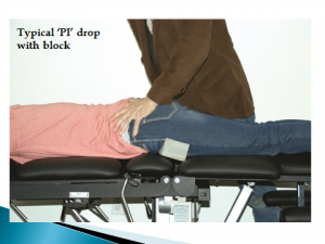
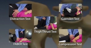
That most orthopedic tests haven’t been proven definitive in diagnosing some or most musculoskeletal conditions is a given. In fact even those tests with good inter-reliability suffer with short comings in validity. Laslett, Bogduk et al did a systematic review in 2010 to determine whether tests could accurately determine a disc vs. facet vs. an SI joint and found the answer to be “generally NO”. However most experts suggest a barrage of tests (with the majority negative (or positive)) give an acceptable odds-ratio as to the likelihood of involvement. This is more so the case with the SI joint vs. facet or disc.

The above picture from SI-Bone suggests a clinical testing procedure with the (5) most diagnostically-relevant SI tests. IF (4) of the (5) are positive there is a 75% likelihood the SI may be involved. The opposite is true as well ((4) of (5) negative). However other studies suggest the MOST relevant tests are the THIGH TRUST and distraction/compression. These (2) tests tended to give the strongest indications.   

Effects of augmented trunk stabilization with external compression support on shoulder & scapular muscle activity and strength during isometric shoulder abduction.

Janng HU et al. J Electromyo Kneiso Apr;25(2) 2014

This study gives more directed research to the phenomena behind the ATM2 restraint/belting/repositioning. The authors wanted to evaluate the ATM2 (external pelvic and/or thoracic compression) effects via EMG on muscle activation properties with external support (the ATM2 premise).  They evaluated several scapular stabilization muscles (upper trapezius, serratus, middle deltoid) under (3) conditions:

  1. NO external support
  2. Pelvic support alone
  3. Both pelvic and shoulder thoracic support. During abduction, strength was viably increased under both pelvic and thoracic compression/restraint (condition (3)).

The findings suggest that: “augmented trunk stabilization may be advantageous with regard to reducing the compensatory muscle efforts of trapezius during isometric shoulder abduction and increasing of abduction strength”.